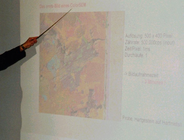
The worldwide first "ColorSEM"-image, presented at a meeting on May 28. 2001. The colored image with a size of 500 x 400 pixel was taken with 3 minutes acqisition time only (500 kcps input!).
(full size
 )
)
The color information is mixed automatically with the electron microscope image during acquisition. The image gives very impressive information about element distributions at specimen surface in addition to the topografic information of common electron image (Element Imaging).
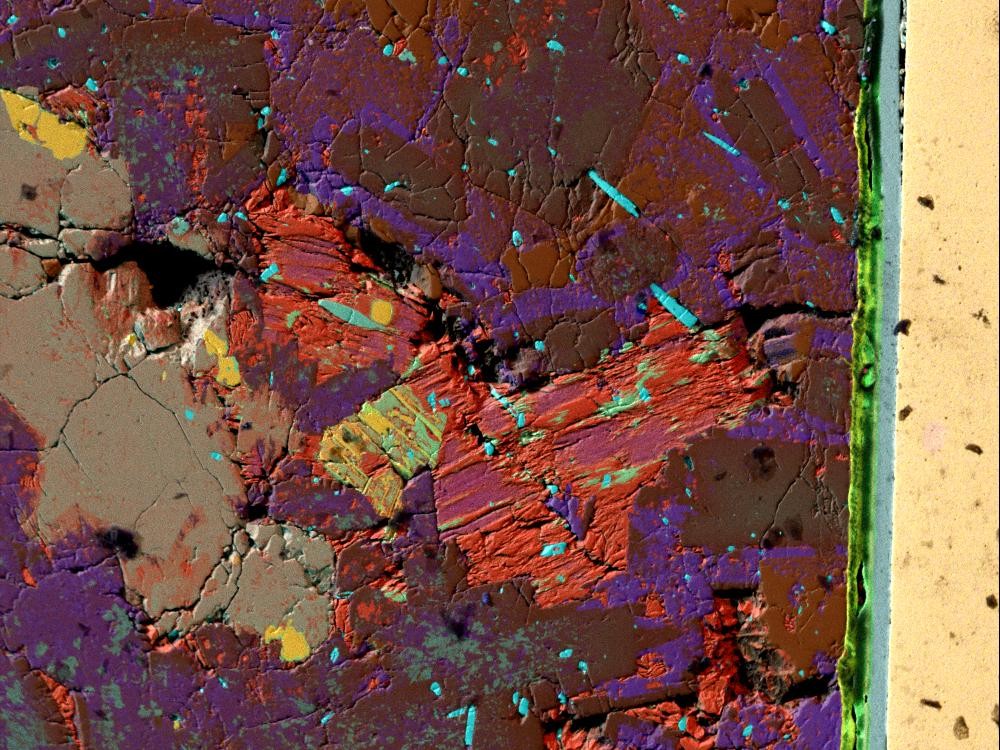
3 years before:
This 'historic' Element Imaging picture of rock cross section was taken at May 1998 in the facilities of Point Electronic company (Halle/Saale, Germany). The acquisition time was 10 minutes, for all element images at same time. 250.000 counts per second (250 kcps!) were processed. These high count rates with Energy Dispersive Detector (EDS, EDX, X-Ray Spectrometer) were possible only with the new working principle of a SDD (Silicon Drift Detector) from KETEK - Company (Munich).
Many thanks to Mr. Joachimi (Point Electronic) for the very successful collaboration in this field for many years and for the very interesting pioneer work in the field of taking element images with X-ray detector in Electron Microscopes with very high count rates (Element Imaging).
back
MICROANALYST.NET